Anatomy Of Musckes Sndctendons / Appendicular Muscles of the Pelvic Girdle and Lower Limbs ... / Tendons are tough bands of dense.. The broad muscle that covers the top of the skull raises eyebrows, wrinkles forehead This review will also illustrate the vascular and lymphatic network and the innervating nerve branch. Smooth muscle contractions are involuntary movements triggered by. Muscular contraction is necessary for voluntary and involuntary movement of limbs, stabilization of joints, maintaining luminal diameter (in the case of arteries, bowel, etc), and to produce heat. Home > blog > anatomy > shoulder anatomy:
Muscle mass accounts for a large majority of the carcass weight of domestic animals. Attached to the bones of the skeletal system are about 700 named muscles that make up roughly half of a person's body weight. Along with lateral pterygoid muscle it produces side to side movement of mandible. The muscles of the torso, examined in the previous chapter, include a few that attach directly into the upper arm and help move the humerus at the shoulder joint. Understanding the structure of a muscle fiber.
Gastrocnemius & Soleus (Calf) muscles from www.musclesused.com It elevates and protrudes the mandible. A collection of anatomy notes covering the key anatomy concepts that medical students need to learn. Attached to the bones of the skeletal system are about 700 named muscles that make up roughly half of a person's body weight. Understanding the structure of a muscle fiber. Muscular contraction is necessary for voluntary and involuntary movement of limbs, stabilization of joints, maintaining luminal diameter (in the case of arteries, bowel, etc), and to produce heat. Seventeen muscles attach to the scapula, and it articulates with the clavicle to form the shoulder girdle or pectoral girdle, which supports movements. Knee function is determined in large part by the anatomy of the joint. Learn about the muscles, tendons, bones, and ligaments that comprise the knee joint anatomy.
Roll your mouse over any muscle in the diagram below to learn its name.
Lesson on the anatomy of the forearm: Skeletal muscles allow the body to move and maintain posture; The interactive muscle anatomy diagram shown below outlines the major superficial (i.e. Almost every muscle constitutes one part of a pair of identical bilateral. There's no strict demarcation or dividing line between the tendon and the covering around this muscle but that covering is called is called the epimysium fp my cm and it's really just connective tissue that covers the muscle kind of protects it reduces friction. Anatomical terms structures of the knee bones of the knee ligaments in the knee cartilage of the knee muscles around the knee tendons in the there are numerous tendons around the knee that also help to stabilize the knee. Muscles of mastication are classified as main and accessory muscles. Along with lateral pterygoid muscle it produces side to side movement of mandible. Specifically, the four rotator cuff muscles. In the muscular system, muscle tissue is categorized into three distinct types: Learn anatomy faster and remember everything you learn. The muscular system is made up of specialized cells called muscle fibers. Knee function is determined in large part by the anatomy of the joint.
Anatomy of the short head of the biceps brachii muscle. Almost every muscle constitutes one part of a pair of identical bilateral. However, if you take a little time to learn a few root words, those latin names can give you valuable insights into things like the muscle's size and shape. Anterior muscles of the neck. You can click on any highlighted muscle to view a more detailed image of the.
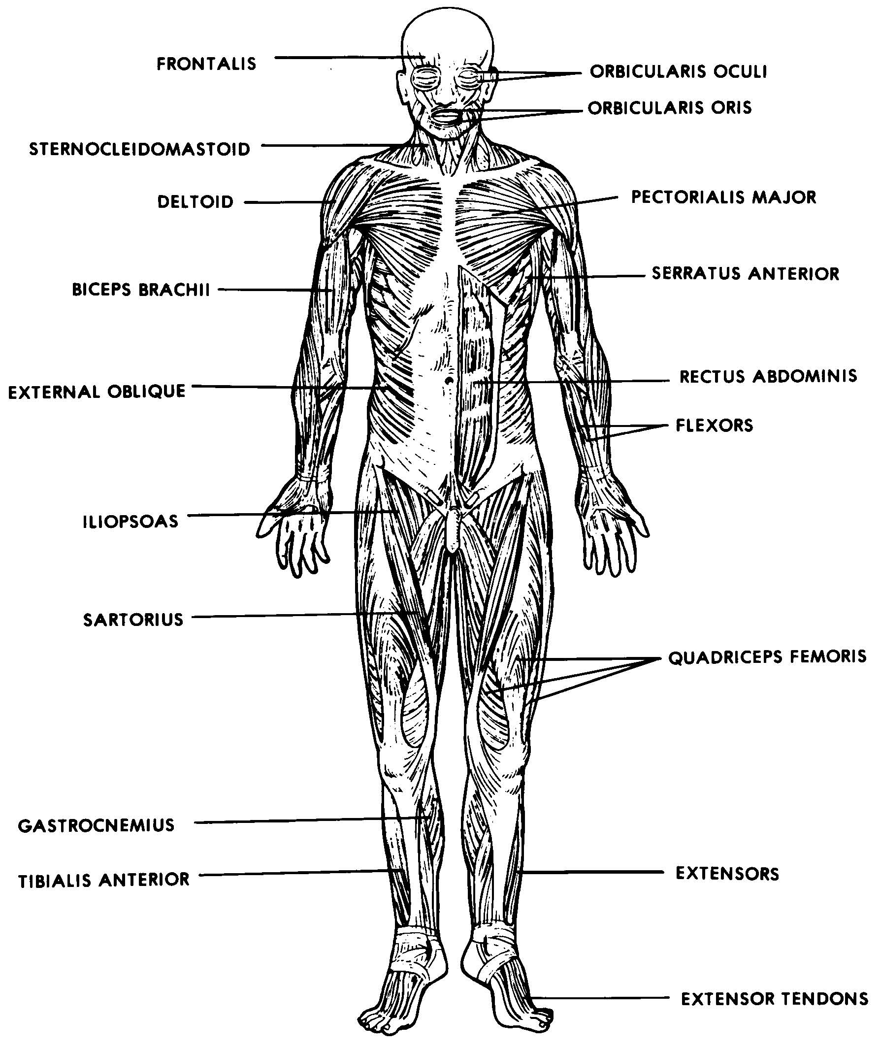
Images 05. Muscular System | Basic Human Anatomy from brooksidepress.org They are associated with muscles discussed in the section above (see. But muscle is also the dominant tissue in the heart and in the walls of other hollow organs of the body. This article reviews the anatomical and functional information of the gastrocnemius muscle, its embryological derivation. Anterior muscles of the neck. Rectus capitis, longus capitis, longus colli. Sternohyoid, sternothyroid, thyrohyoid, omohyoid anterior vertebral muscles: All about the shoulder muscles. The tendons of these muscles pass through a small corridor in the wrist known as the carpal tunnel.
The text describes the concept of fascial continuum, which explains the.
The text describes the concept of fascial continuum, which explains the. Learn about the muscles, tendons, bones, and ligaments that comprise the knee joint anatomy. This review will also illustrate the vascular and lymphatic network and the innervating nerve branch. This article will focus on tongue embryology, origin, insertion, and action of the extrinsic muscles, followed by innervation, blood supply and lymphatic drainage of the tongue. Along with lateral pterygoid muscle it produces side to side movement of mandible. Knee function is determined in large part by the anatomy of the joint. Anatomical terms structures of the knee bones of the knee ligaments in the knee cartilage of the knee muscles around the knee tendons in the there are numerous tendons around the knee that also help to stabilize the knee. Specifically, the four rotator cuff muscles. There's no strict demarcation or dividing line between the tendon and the covering around this muscle but that covering is called is called the epimysium fp my cm and it's really just connective tissue that covers the muscle kind of protects it reduces friction. Seventeen muscles attach to the scapula, and it articulates with the clavicle to form the shoulder girdle or pectoral girdle, which supports movements. Anatomy of the short head of the biceps brachii muscle. Attached to the bones of the skeletal system are about 700 named muscles that gross anatomy of a skeletal muscle most skeletal muscles are attached to two bones through tendons. Digastric, mylohyoid, geniohyoid, stylohyoid infrahyoid muscles:
Learn about the muscles, tendons, bones, and ligaments that comprise the knee joint anatomy. Convergent muscles contain fibers that have a wide origin, but converge in order to attach to a narrow tendon. Anterior muscles of the neck. Topographically, the muscles in this group are classed along with the lateral torso wall and upper extremity , which is due to their location as well as their genetic development based on their embryological origin. Specifically, the four rotator cuff muscles.
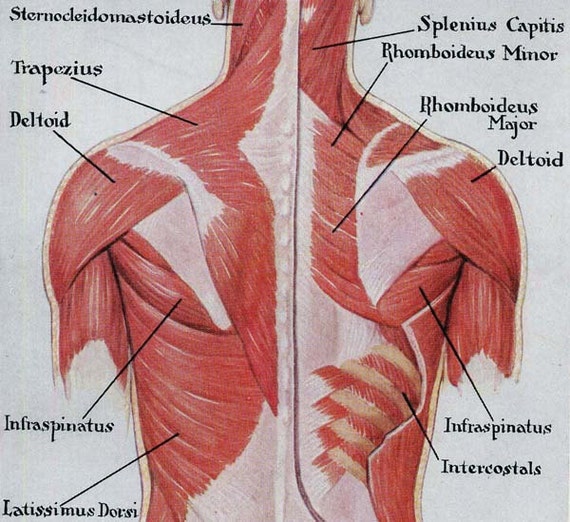
Muscles Back Posterior Human Anatomy Vintage Medical Chart from img1.etsystatic.com Digastric, mylohyoid, geniohyoid, stylohyoid infrahyoid muscles: You can click on any highlighted muscle to view a more detailed image of the. This is a table of skeletal muscles of the human anatomy. There's no strict demarcation or dividing line between the tendon and the covering around this muscle but that covering is called is called the epimysium fp my cm and it's really just connective tissue that covers the muscle kind of protects it reduces friction. Convergent muscles contain fibers that have a wide origin, but converge in order to attach to a narrow tendon. Understanding the structure of a muscle fiber. In the muscular system, muscle tissue is categorized into three distinct types: Anatomy of the short head of the biceps brachii muscle.
This article will focus on tongue embryology, origin, insertion, and action of the extrinsic muscles, followed by innervation, blood supply and lymphatic drainage of the tongue.
This article will focus on tongue embryology, origin, insertion, and action of the extrinsic muscles, followed by innervation, blood supply and lymphatic drainage of the tongue. Rectus capitis, longus capitis, longus colli. The muscles of mastication are a group of muscles associated with movements of the jaw. What of anatomy an essential textbook. How to study muscle anatomy. Convergent muscles contain fibers that have a wide origin, but converge in order to attach to a narrow tendon. Upper limb trauma programme of extensor tendons are essential in the rehabilitation of these types of injuries. This review will also illustrate the vascular and lymphatic network and the innervating nerve branch. Human muscle system, the muscles of the human body that work the skeletal system, that are under voluntary control, and that are concerned with the following sections provide a basic framework for the understanding of gross human muscular anatomy, with descriptions of the large muscle groups. The text describes the concept of fascial continuum, which explains the. They are associated with muscles discussed in the section above (see. Understanding the structure of a muscle fiber. Inflammation of this region caused by repetitive stress or trauma may lead to pain and numbness known as carpal tunnel syndrome.


0 Komentar